

What Are The Three Different Types Of Muscle Tissue Found In Animals?
| Muscle tissue | |
|---|---|
 The body contains iii types of muscle tissue: (a) skeletal muscle, (b) smooth muscle, and (c) cardiac musculus. (Same magnification) | |
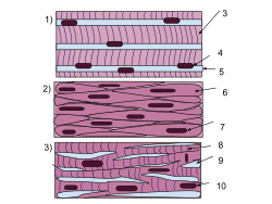 A schematic diagram of the different types of muscle cells (aforementioned guild as above). | |
| Anatomical terminology [edit on Wikidata] |
Muscle tissues are soft tissues that make up the dissimilar types of muscles in most animals, and give the power of muscles to contract. It is also referred to equally myopropulsive tissue. Muscle tissue is formed during embryonic evolution, in a procedure known as myogenesis. Muscle tissue contains special contractile proteins chosen actin and myosin which contract and relax to cause movement. Amongst many other muscle proteins nowadays are two regulatory proteins, troponin and tropomyosin.
Muscle tissues vary with role and location in the torso. In mammals the three types are: skeletal or striated muscle tissue; smooth muscle (non-striated) musculus; and cardiac muscle. Skeletal muscle tissue consists of elongated muscle cells chosen muscle fibers, and is responsible for movements of the trunk. Other tissues in skeletal muscle include tendons and perimysium.[ane] Smooth and cardiac muscle contract involuntarily, without conscious intervention. These muscle types may exist activated both through the interaction of the central nervous organization as well as by receiving innervation from peripheral plexus or endocrine (hormonal) activation. Striated or skeletal muscle only contracts voluntarily, upon the influence of the fundamental nervous system. Reflexes are a form of non conscious activation of skeletal muscles, but however arise through activation of the central nervous system, albeit not engaging cortical structures until afterward the contraction has occurred.[1]
The unlike muscle types vary in their response to neurotransmitters and hormones such as acetylcholine, noradrenaline, adrenaline, and nitric oxide depending on musculus type and the verbal location of the muscle.[1]
Sub-categorization of muscle tissue is too possible, depending on amid other things the content of myoglobin, mitochondria, and myosin ATPase etc.[ commendation needed ]
Structure [edit]

Three distinct types of muscle (L to R): Smooth (non-striated) muscle in internal organs, cardiac or heart musculus, and skeletal muscle.
In that location are three types of muscle tissue in vertebrates: skeletal, cardiac, and smooth. Skeletal and cardiac muscle are types of striated musculus tissue.[2] Shine musculus is non-striated.
Skeletal muscle tissue is an elongated striated musculus tissue ranging from several millimeters to about ten centimeters in length and from x to 100 micrometers in width.[3] Skeletal striated muscle tissue is bundled in regular, parallel bundles of myofibrils containing the many contractile units known equally sarcomeres, which requite the tissue its striated (striped) appearance. Skeletal muscle, is voluntary musculus anchored by tendons or sometimes by aponeuroses to bones, and is used to effect skeletal movement such as locomotion and to maintain posture. Postural control is generally maintained as an unconscious reflex, just the muscles responsible tin can likewise react to witting command. An average adult man is fabricated upward of 42% of skeletal muscle as a percent of body mass, and an boilerplate adult woman is made upwards of 36%.[4]
Cardiac muscle tissue, is plant only in the walls of the heart as myocardium, and is involuntary existence controlled past the autonomic nervous system. Cardiac muscle tissue is striated similar skeletal musculus, containing contractile units called sarcomeres in highly regular arrangements of bundles. While skeletal muscles are arranged in regular, parallel bundles, cardiac muscle connects at branching, irregular angles known as intercalated discs.
Smoothen musculus tissue is non-striated and involuntary. Smoothen muscle is found inside the walls of organs and structures such as the esophagus, stomach, intestines, bronchi, uterus, urethra, bladder, claret vessels, and the arrector pili in the skin which controls the erection of trunk hair.
Comparison of types [edit]
| smooth muscle | cardiac muscle | skeletal muscle | |
| Anatomy | |||
| Neuromuscular junction | none | nowadays | |
| Fibers | fusiform, short (<0.iv mm) | branching | cylindrical, long (<15 cm) |
| Mitochondria | numerous | many to few (by type) | |
| Nuclei | i | 1 | >1 |
| Sarcomeres | none | nowadays, max. length two.vi µm | present, max. length 3.7 µm |
| Syncytium | none (independent cells) | none (but functional as such) | present |
| Sarcoplasmic reticulum | little elaborated | moderately elaborated | highly elaborated |
| ATPase | little | moderate | abundant |
| Physiology | |||
| Self-regulation | spontaneous activity (slow) | yes (rapid) | none (requires nerve stimulus) |
| Response to stimulus | unresponsive | "all-or-zippo" | "all-or-cipher" |
| Action potential | yes | yep | yes |
| Workspace | Force/length bend is variable | the increment in the force/length bend | at the tiptop of the forcefulness/length bend |
| Response to stimulus |  |  |  |
Skeletal muscle [edit]
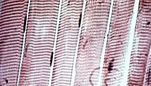
Striated skeletal muscle cells in microscopic view. The myofibers are the direct vertical bands; the horizontal striations (lighter and darker bands) that are a visible consequence from differences in limerick and density along the fibrils within the cells. The cigar-similar dark patches beside the myofibers are muscle-jail cell nuclei.
Skeletal musculus is broadly classified into two cobweb types: Type I tiresome-twitch, and Type Ii fast-twitch muscle.
- Type I, slow-twitch, ho-hum oxidative, or red muscle is dumbo with capillaries and is rich in mitochondria and myoglobin, giving the musculus tissue its characteristic ruddy color. It can comport more oxygen and sustain aerobic activity.
- Type Two, fast-twitch muscle, has three major kinds that are, in lodge of increasing contractile speed:[6]
- Type IIa, which, similar a slow muscle, is aerobic, rich in mitochondria and capillaries and appears red when deoxygenated.
- Type IIx (as well known as blazon IId), which is less dumbo in mitochondria and myoglobin. This is the fastest musculus type in humans. It can contract more chop-chop and with a greater amount of strength than oxidative muscle but can sustain but short, anaerobic bursts of activity before muscle contraction becomes painful (ofttimes incorrectly attributed to a build-upward of lactic acrid). Northward.B. in some books and articles this muscle in humans was, confusingly, called type IIB.[7]
- Blazon IIb, which is anaerobic, glycolytic, "white" muscle that is fifty-fifty less dense in mitochondria and myoglobin. In small animals like rodents, this is the major fast musculus type, explaining the pale colour of their mankind.
The density of mammalian skeletal muscle tissue is near ane.06 kg/liter.[8] This can be contrasted with the density of adipose tissue (fatty), which is 0.9196 kg/liter.[9] This makes muscle tissue approximately 15% denser than fatty tissue.
Smooth musculus [edit]
Smooth muscle is involuntary and not-striated. Information technology is divided into two subgroups: the single-unit (unitary) and multiunit shine muscle. Inside unmarried-unit cells, the whole package or sheet contracts as a syncytium (i.eastward. a multinucleate mass of cytoplasm that is not separated into cells). Multiunit shine muscle tissues innervate individual cells; as such, they let for fine command and gradual responses, much like motor unit recruitment in skeletal muscle.
Smooth musculus is found inside the walls of blood vessels (such smooth muscle specifically existence termed vascular smooth muscle) such as in the tunica media layer of large (aorta) and small arteries, arterioles and veins. Smooth muscle is besides found in lymphatic vessels, the urinary bladder, uterus (termed uterine smooth muscle), male person and female reproductive tracts, alimentary canal, respiratory tract, arrector pili of peel, the ciliary musculus, and iris of the middle. The structure and function is basically the same in smooth musculus cells in different organs, just the inducing stimuli differ essentially, in club to perform individual effects in the torso at individual times. In addition, the glomeruli of the kidneys contain smooth musculus-similar cells called mesangial cells.
Cardiac muscle [edit]
Cardiac muscle is involuntary, striated muscle that is found in the walls and histological foundation of the centre, specifically the myocardium. The cardiac muscle cells, (also called cardiomyocytes or myocardiocytes), predominantly contain merely ane nucleus, although populations with two to four nuclei practise exist.[ten] [11] [ page needed ] The myocardium is the muscle tissue of the heart and forms a thick middle layer between the outer epicardium layer and the inner endocardium layer.
Coordinated contractions of cardiac muscle cells in the eye propel blood out of the atria and ventricles to the blood vessels of the left/body/systemic and correct/lungs/pulmonary circulatory systems. This complex mechanism illustrates systole of the heart.
Cardiac muscle cells, unlike well-nigh other tissues in the body, rely on an bachelor blood and electrical supply to evangelize oxygen and nutrients and remove waste products such as carbon dioxide. The coronary arteries assistance fulfill this function.
Evolution [edit]
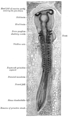
A chicken embryo, showing the paraxial mesoderm on both sides of the neural fold. The anterior (forward) portion has begun to form somites (labeled "archaic segments").
All muscles are derived from paraxial mesoderm. The paraxial mesoderm is divided along the embryo's length into somites, corresponding to the segmentation of the body (nearly plainly seen in the vertebral column.[12] Each somite has three divisions, sclerotome (which forms vertebrae), dermatome (which forms skin), and myotome (which forms muscle). The myotome is divided into two sections, the epimere and hypomere, which form epaxial and hypaxial muscles, respectively. The only epaxial muscles in humans are the erector spinae and small-scale intervertebral muscles, and are innervated by the dorsal rami of the spinal nerves. All other muscles, including those of the limbs are hypaxial, and innervated by the ventral rami of the spinal nerves.[12]
During development, myoblasts (musculus progenitor cells) either remain in the somite to form muscles associated with the vertebral column or drift out into the body to course all other muscles. Myoblast migration is preceded by the formation of connective tissue frameworks, ordinarily formed from the somatic lateral plate mesoderm. Myoblasts follow chemic signals to the appropriate locations, where they fuse into elongate skeletal muscle cells.[12]
Function [edit]
The primary office of muscle tissue is contraction. The three types of musculus tissue (skeletal, cardiac and smooth) take significant differences. Yet, all three utilize the movement of actin against myosin to create contraction.
Skeletal musculus [edit]
In skeletal muscle, contraction is stimulated past electrical impulses transmitted by the motor nerves. Cardiac and smooth musculus contractions are stimulated by internal pacemaker cells which regularly contract, and propagate contractions to other muscle cells they are in contact with. All skeletal musculus and many smooth muscle contractions are facilitated by the neurotransmitter acetylcholine.
Smooth muscle [edit]
Smoothen musculus is found in almost all organ systems such as hollow organs including the stomach, and float; in tubular structures such as blood and lymph vessels, and bile ducts; in sphincters such as in the uterus, and the eye. In addition, it plays an important part in the ducts of exocrine glands. Information technology fulfills diverse tasks such every bit sealing orifices (e.grand. pylorus, uterine os) or the ship of the chyme through wavelike contractions of the abdominal tube. Smooth muscle cells contract more slowly than skeletal muscle cells, but they are stronger, more sustained and crave less energy. Smooth muscle is as well involuntary, unlike skeletal muscle, which requires a stimulus.
Cardiac muscle [edit]
Cardiac muscle is the musculus of the heart. Information technology is self-contracting, autonomically regulated and must go on to contract in a rhythmic fashion for the whole life of the organism. Hence it has special features.
References [edit]
- ^ a b c "Muscle Tissue TheVisualMD.com". www.thevisualmd.com . Retrieved 2015-12-30 .
- ^ Pratt, Rebecca. "Musculus Tissue". AnatomyOne. Amirsys, Inc. Archived from the original on 2 February 2017. Retrieved 26 Oct 2012.
- ^ Hugh Potter, Summary of muscle tissue "Archived copy". Archived from the original on 2014-ten-21. Retrieved 2014-09-02 .
{{cite spider web}}: CS1 maint: archived copy as title (link) CS1 maint: bot: original URL condition unknown (link) - ^ Marieb, Elaine; Hoehn, Katja (2007). Human Anatomy & Physiology (7th ed.). Pearson Benjamin Cummings. p. 317. ISBN978-0-8053-5387-7.
- ^ Talbot, J; Maves, L (July 2016). "Skeletal muscle fiber type: using insights from muscle developmental biology to dissect targets for susceptibility and resistance to muscle disease". Wiley Interdisciplinary Reviews. Developmental Biology. 5 (4): 518–34. doi:10.1002/wdev.230. PMC5180455. PMID 27199166.
- ^ Smerdu, V; Karsch-Mizrachi, I; Campione, One thousand; Leinwand, L; Schiaffino, Southward (December 1994). "Type IIx myosin heavy chain transcripts are expressed in type IIb fibers of human skeletal muscle". The American Journal of Physiology. 267 (6 pt i): C1723–1728. doi:x.1152/ajpcell.1994.267.half dozen.C1723. PMID 7545970. Annotation: Access to total text requires subscription; abstract freely bachelor
- ^ Urbancheka, K; Picken, Eastward; Kalliainen, L; Kuzon, W (2001). "Specific Force Deficit in Skeletal Muscles of Former Rats Is Partially Explained past the Existence of Denervated Musculus Fibers". The Journals of Gerontology Serial A: Biological Sciences and Medical Sciences. 56 (5): B191–B197. doi:10.1093/gerona/56.v.B191. PMID 11320099.
- ^ Farvid, MS; Ng, TW; Chan, DC; Barrett, PH; Watts, GF (2005). "Clan of adiponectin and resistin with adipose tissue compartments, insulin resistance and dyslipidaemia". Diabetes, Obesity & Metabolism. 7 (four): 406–413. doi:10.1111/j.1463-1326.2004.00410.10. PMID 15955127. S2CID 46736884.
- ^ Olivetti G, Cigola Eastward, Maestri R, et al. (July 1996). "Aging, cardiac hypertrophy and ischemic cardiomyopathy do non affect the proportion of mononucleated and multinucleated myocytes in the human heart". Journal of Molecular and Cellular Cardiology. 28 (7): 1463–77. doi:ten.1006/jmcc.1996.0137. PMID 8841934.
- ^ Pollard, Thomas D. and Earnshaw, William. C., "Cell Biology". Philadelphia: Saunders. 2007.
- ^ a b c Sweeney, Lauren (1997). Basic Concepts in Embryology: A Educatee's Survival Guide (1st Paperback ed.). McGraw-Loma Professional person.
Source: https://en.wikipedia.org/wiki/Muscle_tissue
Posted by: goodefifery.blogspot.com
0 Response to "What Are The Three Different Types Of Muscle Tissue Found In Animals?"
Post a Comment